RealityCapture
Creating a virtual atlas of neuroanatomy
Neuroanatomy education has been historically focused on 2D and, more recently, 3D imaging. When available, training with cadavers in the laboratory notably improves the understanding of such challenging anatomy. Unfortunately, cadaveric laboratories are not available worldwide and the high cost of the specimen makes this exercise confined to few residents/neurosurgeons.
Nowadays with the advance in technologies, the use of volumetric models (VMs) from DICOM or illustrations imaging is gaining more popularity in the educational anatomical field. In neurosurgical learning, these VMs are a useful tool to improve understanding of the 3D relationship of the neurovascular structures and to overpass the lack of cadaveric dissections. For these reasons, I decided to concentrate my Ph.D. research (University of Ferrara, Italy) on creating a virtual atlas of neuroanatomy and neurosurgical techniques.
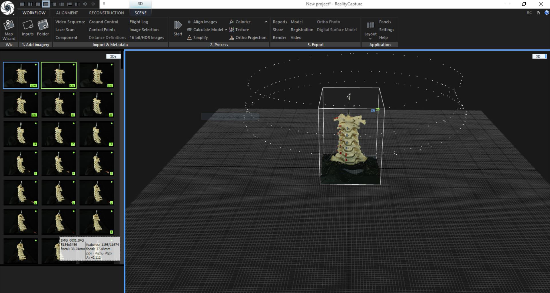
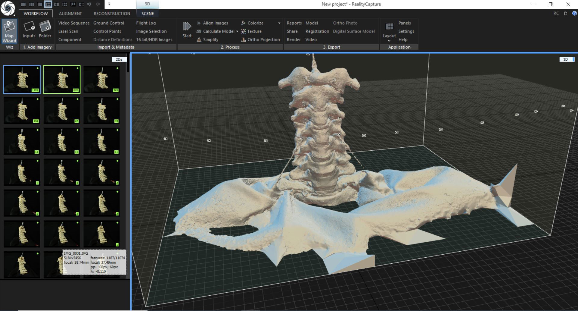
The photogrammetry (PGM) technique to create VM is low cost and easy to perform. The PGM workflow involves only 3 steps: image capture, preprocessing and postprocessing. The advantage of PMG includes the possibility of using different types of cameras such as digital, single-lens reflex, charged couple device, mirrorless camera, smartphone camera. Moreover, the use of surgical devices such as microscopes or endoscopes allows us to perform the reconstruction of VMs of deep anatomical surfaces which, since the small dimension of the neurovascular structures (around millimeter or submillimeter) would be very hard to expose and to detect with other cameras or scanning techniques.
In my experience, Reality Capture is the photogrammetry software that offers the best reconstructions of fine anatomical dissections in an incredibly short time. Creating a model with 200-300 images, like I usually do, takes no more than 15-20 mins, which is far below compared to other programs. This advantage makes the workflow smoother and allows faster pursuance of the project. Moreover, RC has many tools to play with the model during its acquisition and leads to a higher resolution of the mesh and texture of the models.
When a VM is ready I usually upload it to a 3D viewing platform, like Sketchfab, which allows 2D, 3D and extended reality interaction of the models. With Sketchfab is possible to add descriptions and annotations to underline the important structures of each model. These models can be also used to do renders and to create 3D videos that can be incorporate in surgical videos to better understand anatomy.
Moreover, these VMs can be 3D printed which can be a valid substitute when bones or cadaveric specimens are not available. The final purpose is to create a free collection available online where trainees, students, and surgeons can see, play and download these VMs to improve and facilitate neuroanatomy learning.
Article provided by Vera Vigo, University of Ferrara, Italy.